- This event has passed.
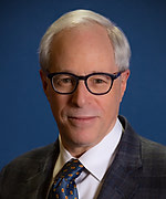
Richard Levenson, MD, UC Davis Health – “Novel Tissue Imaging for Faster Diagnostics, Better AI”
April 4, 2023 @ 10:00 am - 11:00 am
 Light microscopy of tissue biopsies remains the central technique in diagnosis and management of cancers as well as other diseases. The brightfield (transmission) optical design of today’s clinical microscopes requires optically thin slices of tissue mounted onto glass slides, a labor- and time-intensive process. Moreover, tissue sectioning can partially or completely consume small specimens, leaving little or nothing for downstream molecular studies. Separately, when conventional slides are prepared, there are still advances in microscopy techniques than can acquire more and better information as compared to standard slide-viewing or scanning.
Our laboratory is developing novel approaches to histopathology: MUSE, FIBI and DUET. 1) MUSE (based on UV light excitation) and FIBI (a visible-light based approach) are non-destructive techniques that capture high-resolution microscopy images directly from the face of unsectioned tissue specimens. 2) DUET (dual-mode emission-transmission) is designed for use with existing H&E-stained slides, and generates both brightfield and long-pass fluorescence data. A variety of parametric and machine-learning methods can then be used to display collagen, elastin and basement membrane distribution, as well as other components not easily visualized by brightfield-only methods, all without requiring the preparation of additional, alternatively stained tissue sections.
With MUSE, FIBI and DUET time and money can be saved, but in addition, enhanced tissue contrast could drive enhanced performance for computational approaches. In the meantime, slide-free imaging can provide near-patient, near-real-time histology results to monitor of biopsy quality, provide frozen-section replacements for intraoperative surgical guidance, and allow point-of-care tissue diagnostics for remote and/or low-resource settings.
Richard Levenson, MD, FCAP, is Professor and Vice Chair for Strategic Technologies in the Department of Pathology and Laboratory Medicine at UC Davis. Dr. Levenson has helped develop multispectral microscopy and small-animal imaging systems and software, birefringence microscopy, multiplexed ion-beam imaging (MIBI), and most recently, slide-free as well as enhanced-content microscopy approaches. He is section editor for Archives of Pathology and is on the editorial board of Lab, Invest, and AJP. Regrettably, he also taught pigeons histopathology and radiology. He is a recipient of the 2018 UC Davis Chancellor’s Innovator of the Year award and is a Fellow of SPIE.
Note this is a hybrid event, in-person at Wolf Conference Center, 3rd floor of Hale BTM, 60 Fenwood Road and on Zoom.